Intra Oral Dental Imaging Intra Oral Dental Imaging Intra Oral Dental Imaging Intra Oral Dental Imaging Intra Oral Dental Imaging
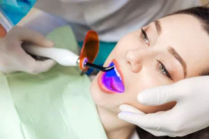
Technology in the dental field has been evolving over time, helping both the dentist and the patients obtain better diagnoses in less time and within everyone’s reach. Thus an adequate treatment plan for patients is possible. As a small cavity, the mouth sometimes prevents adequate visualization of all its structures. In many cases, clinical evaluation is not enough to reach an accurate diagnosis, and it becomes necessary to use auxiliary tests such as radiographs or other methods for obtaining intraoral dental images.
[scroll_to title=”#1″ bullet=”false”]
Menu of Contents
ToggleINTRAORAL RADIOGRAPHS
The vast majority of dental images are created from the application of X-ray technology; to these devices, we can obtain the necessary information to determine a timely and accurate diagnosis. Intraoral radiographs use radiographic films placed inside the oral cavity. There are different types of radiographs, among them we have the following:
- Periapical radiography:
This type of radiography is one of the most requested. It is also known as dental radiography because thanks to it, you can observe the entire tooth from the tip of the root to the crown, which is the visible part in the mouth. From this radiographic image,you can evaluate the state of the tooth and its adjacent structures such as the bone around it and the supporting tissues. It is useful to determine the progress of tooth decay, the presence of pulp involvement, the presence and progress of any infection and to rule out any fracture or periodontal disease.
[row]
[col span__sm=”12″ align=”center”]
[ux_image id=”22132″ width=”55″]
[ux_image id=”20080″ width=”36″]
[/col]
[/row]
- Bitewing or interproximal radiographs:
These are radiographs of the same size as the periapical type used with a different technique to obtain the intraoral images that will help us obtain information about a greater number of teeth, but only of the coronary portion of both dental arches; that is to say, of the upper and lower jaw. One of the main reasons for requesting this type of radiography is to determine the presence of dental caries between teeth, which is often difficult to diagnose in a clinical examination. It is also useful for the evaluation of the presence and progression of disease of the supporting structures (periodontium). Intra Oral Dental Imaging
- Occlusal Radiography:
These radiographs are slightly larger compared to periapical radiographs. In order to obtain this intraoral image, a technique is used in which the patient bites the radiographic plate. With the radiograph, it is possible to evaluate the location of teeth that have not yet erupted in the mouth or those in a different position than normal.
scroll_to title=”#2″ bullet=”false”]
EXTRAORAL RADIOGRAPHS
[row]
[col span__sm=”12″ align=”center”]
[ux_image id=”22132″ width=”55″]
[ux_image id=”20078″ width=”39″]
[/col]
[/row]
Intraoral radiographs are not the only way to obtain dental images. There are also extraoral radiographs. Itt is already very common to obtain images through the use of cone beam computed tomography (CBTC); this type of image generates more information because the image is in different planes as a three-dimensional image (3D). An investigation published in the journal of oral and maxillofacial surgery, conducted by the academic and professional community of WHO determined that the use of cone beam tomography is safe since it ensures safety parameters for the patient in terms of radiation exposure. Intra Oral Dental Imaging
The decision of what type of image to request depends on different factors but fundamentally it is based on what is to be observed: the position of a tooth, the presence of periodontal disease, fractures, infections, the evaluation of any treatment, among others. It is important to emphasize that dental x-ray examinations are safe according to the American Dental Association (ADA) since they have a very low level of radiation exposure, which makes the risk of possible harmful effects very small. Intra Oral Dental Imaging
[scroll_to title=”#3″ bullet=”false”]
INTRAORAL SCANNING
This type of intraoral imaging allows us to create a 3D digital file of the patient’s mouth and thus be able to elaborate the work digitally, reducing processing time often affording greater accuracy. This system will replace the traditional models made from the patient’s mouth, giving greater comfort to the patient.
[scroll_to title=”4″ bullet=”false”]
CONTACT US
If you have any questions about this topic or any other, please feel free to contact or visit us at Channel Island Family Dental. As well as on our Facebook page. At Channel Island Family Dental, we are always attentive to your needs to make a timely diagnosis. In addition, our dentists in Oxnard, Santa Paula, Newbury Park Ventura, and Port Hueneme will guide you to the best treatment to give you back your best smile.
Intra Oral Dental Imaging Intra Oral Dental Imaging Intra Oral Dental Imaging Intra Oral Dental Imaging Intra Oral Dental Imaging